 Partner with Us
  Referring Provider Portal
  Patients: Request Appointment
  Contact Us


Retina Care And Treatment
Damage to the retina can permanently affect sight if it goes untreated. The retina is the tissue at the back of the eye that makes vision possible.
 AVP•SEES Group retina specialists diagnose and treat conditions such as retinal detachment, diabetic retinopathy, macular degeneration, and retinal tears. We serve patients across Virginia, Tennessee, and Alabama, using advanced imaging to protect and preserve long-term vision.
Retina Care at AVP•SEES Group
Clear vision depends on more than a healthy lens and cornea. At the back of the eye, a thin layer of tissue called the retina converts light into signals. Your brain uses those signals to create the images you see.
Damage anywhere in that layer, even in a small area, can change how well you see. AVP•SEES Group retina specialists evaluate and treat these changes for patients across Virginia, Tennessee, and Alabama. We combine detailed imaging with hands-on clinical experience to guide each patient’s care.
Symptoms of Retinal Disease
Some retinal problems build slowly over years. Others show up overnight. Because early-stage retinal disease produces no warning signs, an eye exam is often the only way to catch it early. This matters most for people managing diabetes or high blood pressure. It also applies to anyone with a parent or sibling who has had retinal disease, or anyone at higher risk because of age.
Watch for any of the following:
- Vision that looks blurry or out of focus
- Missing patches or dark spots within your view
- Quick flashes of light, especially in low light
- A rise in the number of floaters you notice
- A curtain-like shadow moving across part of your sight
- Trouble making out fine print or familiar faces
- A drop in either your central or side vision
- Straight lines that appear bent or wavy
Vision changes are not always related to the retina. When symptoms point to the brain or optic nerve instead, our neurological eye conditions team evaluates those concerns separately.
Retinal Conditions We Treat
The conditions our retina team manages range from slow, chronic disease to sudden, sight-threatening emergencies.
- Diabetic retinopathy
- Age-related macular degeneration (AMD)
- Retinal tears and detachment
- Vein and artery occlusion in the retina
- Swelling of the macula (macular edema)
- Scar tissue on the retina (epiretinal membrane)
- Bleeding within the eye (vitreous hemorrhage)
- Inflammatory conditions affecting the retina
- Disease affecting the outer edges of the retina (peripheral retinal disease)

Diabetic Retinopathy
Diabetes does not just affect blood sugar. Over time, it can weaken and damage the tiny blood vessels that feed the retina, a condition called diabetic retinopathy. The National Eye Institute identifies it as the leading cause of vision loss among people with
 Trusted Source
Diabetic Retinopathy
National Eye Institute
Go to Source
diabetes.
Early stages often pass without any noticeable symptoms. Left unchecked, advanced cases can also trigger a form of glaucoma or other retinal complications. An annual dilated eye exam remains the most reliable way to catch changes before they progress.
Trusted Source
Diabetic Retinopathy
National Eye Institute
Go to Source
diabetes.
Early stages often pass without any noticeable symptoms. Left unchecked, advanced cases can also trigger a form of glaucoma or other retinal complications. An annual dilated eye exam remains the most reliable way to catch changes before they progress.
Age-Related Macular Degeneration (AMD)
Age-related macular degeneration, or AMD, targets the macula, the small central area of the retina that handles fine detail. This is why many older adults find reading, driving at night, or recognizing a familiar face gradually harder. According to the American Society of Retina Specialists, AMD appears in two
Trusted Source
Age-Related Macular Degeneration
American Society of Retina Specialists
Go to Source
forms.
A slower dry form is more common, while a faster, more aggressive wet form progresses quickly. Which type a patient has shapes the treatment plan.
Retinal Tears and Retinal Detachment
A retinal tear happens when a small opening forms in the retina. If that opening allows the retina to separate from the tissue behind it, the result is a retinal detachment. This is a much more serious problem. Because the retina can no longer relay images to the brain once it separates, the warning signs are usually dramatic. They can include a burst of floaters, flashing lights, blurred vision, or a dark curtain on one side of your view. The National Eye Institute classifies retinal detachment as a medical
Trusted Source
Retinal Detachment
National Eye Institute
Go to Source
emergency.
Getting treated quickly gives patients the best chance at preserving their sight.
Retinal Vein and Artery Occlusion
A retinal vein occlusion happens when a clot blocks one of the veins that drains blood from the retina. According to the National Eye Institute, this most often affects only one
Trusted Source
Central Retinal Vein Occlusion (CRVO)
National Eye Institute
Go to Source
eye.
Some patients notice no symptoms at all, while others develop sudden blurry vision. A retinal artery occlusion is less common but more urgent. It cuts off blood flow to the retina and typically causes sudden, painless vision loss that needs emergency care. Both conditions raise the risk of complications such as macular edema or, in some cases, glaucoma.
Macular Edema
Macular edema happens when fluid builds up inside the macula. This tiny hub of the retina handles your sharpest, most detailed vision. According to the American Society of Retina Specialists, the most effective treatments address the underlying
Trusted Source
Macular Edema
American Society of Retina Specialists
Go to Source
cause,
whether that is diabetic retinopathy, a blocked retinal vein, or inflammation inside the eye. The result is central vision that looks blurred or warped, rather than a total loss of sight. Catching it early matters: injections or a course of laser therapy can bring the swelling down before it causes lasting damage.
Epiretinal Membrane
As the eye ages, the gel-like vitreous inside it can pull slightly away from the retina. Sometimes that process leaves behind a thin film of scar tissue on the retina’s surface, known as an epiretinal membrane. According to the American Society of Retina Specialists, most cases cause minimal symptoms and can be observed over time without
Trusted Source
Epiretinal Membranes
American Society of Retina Specialists
Go to Source
treatment.
When it does distort vision, a vitrectomy, a surgical procedure to remove the scar tissue, is typically the recommended fix.

Comprehensive Retinal Evaluation
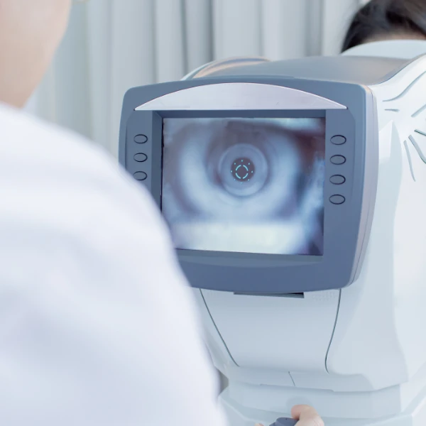
Many retinal diseases progress quietly, without pain or obvious warning signs. That is exactly why imaging plays such a large role in catching them. A clinical exam alone often is not enough.
Our retina specialists rely on a combination of imaging tools to see what the naked eye cannot:
- Optical coherence tomography (OCT): cross-sectional scans that reveal each layer of the retina
- Wide-field retinal imaging: captures the far edges of the retina, where tears often start
- Fundus photography: creates a photographic record for tracking changes visit to visit
- Fluorescein angiography: highlights blood flow through the retina and flags blocked or leaking vessels
- Ocular ultrasound: provides a clear view of the retina when bleeding or clouding blocks a direct look
Retina FAQ
What does a retina specialist do?
Retina specialists complete additional fellowship training beyond general ophthalmology. That training focuses entirely on the retina, macula, vitreous, and the blood vessels behind the eye. This is what lets them diagnose and manage conditions such as diabetic retinopathy, retinal detachment, macular degeneration, and macular edema.
When should I see a retina specialist?
A referral often comes after an eye doctor notices something on an exam, but you do not need to wait. Sudden floaters, flashes of light, or a shadow creeping across your vision are all good reasons to get evaluated directly. The same is true if you already have a diagnosis like diabetic eye disease or macular degeneration.
What are the symptoms of a retinal tear or detachment?
Watch for a spike in floaters, flashes of light, blurry vision, or a shadow that creeps in from one side. Because these can signal a detaching retina, and delaying care risks permanent vision loss, prompt evaluation is the safer path.
What is diabetic retinopathy?
It is the eye complication that comes with long-term diabetes, caused by damage to the blood vessels feeding the retina. Symptoms often do not show up until the disease has already progressed. That is why annual screening matters, even when your vision still feels normal.
What is age-related macular degeneration?
AMD targets the macula, the small central zone of the retina used for detailed tasks. Reading a menu or recognizing a face across the room can become gradually harder. It becomes more common with age, especially after 50, with a slower dry form and a faster wet form.
What tests are used during a retina evaluation?
Your visit may include several imaging steps: OCT scans, wide-field retinal photos, fundus photography, fluorescein angiography, or ocular ultrasound. Which combination you need depends on your symptoms and what your doctor sees during the exam.
Why do patients with diabetes need retina exams?
High blood sugar gradually wears down the small vessels feeding the retina, and that wear can go unnoticed for years. An annual retina exam is how we catch that damage while it remains treatable.

Schedule Your Retina Evaluation
If you are experiencing sudden vision changes, flashes of light, or new floaters, one of our retina specialists can help. The same is true for a diagnosis of diabetic eye disease or macular degeneration. Our team of more than 100 physicians serves patients at locations across Virginia, Tennessee, and Alabama. We bring more than 40 years of combined experience and over 50,000 procedures performed each year.
1 American Society of Retina Specialists — What Is a Retina Specialist? https://www.asrs.org/patients/what-is-a-retina-specialist
2 American Society of Retina Specialists — Age-Related Macular Degeneration. https://www.asrs.org/patients/age-related-macular-degeneration
3 National Eye Institute — Diabetic Retinopathy. https://www.nei.nih.gov/eye-health-information/eye-conditions-and-diseases/diabetic-retinopathy
4 National Eye Institute — Retinal Detachment. https://www.nei.nih.gov/eye-health-information/eye-conditions-and-diseases/retinal-detachment
5 National Eye Institute — Central Retinal Vein Occlusion (CRVO). https://www.nei.nih.gov/eye-health-information/eye-conditions-and-diseases/central-retinal-vein-occlusion-crvo
6 American Society of Retina Specialists — Macular Edema. https://www.asrs.org/patients/retinal-diseases/20/macular-edema
7 American Society of Retina Specialists — Epiretinal Membranes. https://www.asrs.org/patients/retinal-diseases/19/epiretinal-membranes
Leadership at AVP•SEES Group has either authored or reviewed and approved this content.